FIGURE

Fig. 1

ID
ZDB-FIG-210324-19
Publication
Niu et al., 2021 - The Bdkrb2 gene family provides a novel view of viviparity adaptation in Sebastes schlegelii
Other Figures
All Figure Page
Back to All Figure Page
Fig. 1

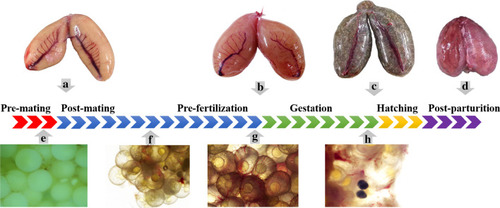
Macroscopic and microcosmic observation of the ovary at different stages of reproductive process. a Ovary at the stage of post-mating. Its overall color is yellow, because of fewer blood vessels in the ovary. b Ovary after fertilization, at the early stage of gestation. Its overall color is pink or red, because the well-developed blood vessels spread over the surface of egg membrane. c Ovary at the final stage of gestation. Its overall color is dark, because of developed pigment. The silvery reflections are the individual eyes of the larvae within the ovary. As time goes by, the ovary will enlarge the volume and the ovarian wall will become very thin until the finish of parturition. d Ovary post parturition. Most embryos came out from cloacal orifice via abdomen extrusion. e Ovary before mating. Oocytes are opaque. Only a few of blood vessels were observed. f Ovary after mating. Oocytes develop further, becoming transparent. g Ovary before fertilization. Oocytes are fully developed and ready for fertilization. The highly vascular membranous tissue adjacent to the egg membrane. h Ovary during gestation. The eyes of embryo formed

Expression Data

Expression Detail
Antibody Labeling
Phenotype Data

Phenotype Detail
Acknowledgments
This image is the copyrighted work of the attributed author or publisher, and ZFIN has permission only to display this image to its users. Additional permissions should be obtained from the applicable author or publisher of the image. Full text @ BMC Ecol Evol