|
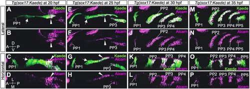
Expression analysis of Alcam in the PP endoderm of Tg(sox17:Kaede). (A-D) At 20 hpf, strong expression of Alcam was evident in C2 but hardly detected in PP1 and R2 endoderm. (E-H) At 25 hpf, Alcam was high in PP3 and the caudal portion of PP2 but almost absent in PP1 and the rostral part of PP2. (I-L) At 30 hpf, high accumulation of Alcam was detected in PP3, PP4 and the caudal layer of PP2, whereas it was at a very low level in PP1 and the rostral layer of PP2. (M-P) At 35 hpf, high accumulation of Alcam was detected in PP3, PP4, PP5 and the caudal part of PP2, whereas it was at a very low level in PP1 and the rostral layer of PP2. A, anterior; D, dorsal; L, lateral; M, medial; P, posterior; PP1-5, the first to fifth pharyngeal pouches; V, ventral. Arrows indicate R2; arrowheads indicate C2. Scale bar: 50 μm. More than two embryos were analyzed at each time point.
|