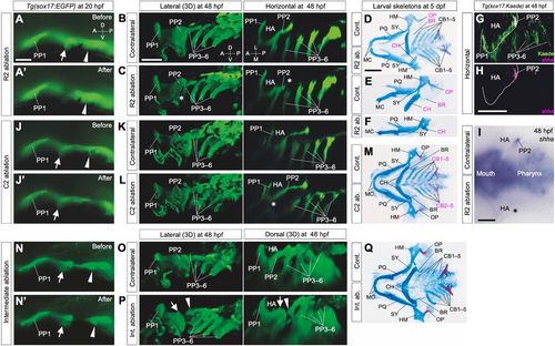
Early determination of distinct roles for later skeletal patterns in R2 and C2 endoderm. (A,A′) Cells of R2 (arrows) in Tg(sox17:EGFP) embryos were ablated at 20 hpf. (B-F) R2 ablation caused a specific loss of epithelial expansion of the caudal lining of the HA (asterisks in C; n=3/3) and reductions in HA-derived skeletal elements, especially in the opercular series (D-F; OP, n=10/16; BR, n=12/16). In addition, this ablation occasionally caused a size reduction in other HA-derived skeletal structures, such as the HM (Fig. S3C; n=3/16) and CH (D-F; n=6/16). (G,H) Expression of shha, required for opercular development, was detected in PP2 endoderm occupied by R2 descendants. (I) Consistent with endodermal (B,C) and the skeletal (D-F) phenotypes, R2 ablation caused a specific loss of shha expression in PP2, as shown in a flat-mounted embryo (asterisk in I; n=12). (J,J′) Cells of C2 (arrowheads) in Tg(sox17:EGFP) embryos were ablated at 20 hpf. (K-M) Ablation of C2 cells resulted in loss of the proximal region of PP2, which consists of the rostral lining of the third PA (BA1) (K,L, asterisk in L; n=3/3), resulting in loss of CB1 cartilage (M; n= 8/8). (N,N′) Endodermal cells between R2 (arrows) and C2 (arrowheads) were ablated in Tg(sox17:EGFP) embryos at 20 hpf. (O-Q) Ablation of cells in the intermediate region did not affect segregation of HA and BA1 but caused abnormal arrangements of them, shown by a split between HA and BA1 (O,P; n=10/12). Correspondingly, on the ablated sides, the positions of BA1-derived CB1 cartilage shifted posteriorly, although a complete set of the pharyngeal structures developed (Q; n=4/6). Images of ablated sides (C,F,L,Q) are inverted in a left-right direction for comparisons with contralateral sides. A, anterior; BR, branchiostegal ray; CB1-5, the first to fifth ceratobranchials; CH, ceratohyal cartilages; D, dorsal; HA, hyoid arch; HM, hyomandibular; L, lateral; M, medial; MC, Meckel's cartilage; OP, opercular bone; P, posterior; PP1-6, the first to sixth pharyngeal pouches; PQ, palatoquadrate; SY, symplectic; V, ventral. Scale bars: 20 μm (A), 50 μm (B,H,I) and 100 μm (D).
|
