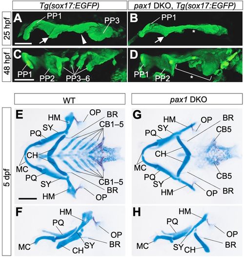
Pharyngeal abnormalities in pax1 dKO mutant embryos. (A-D) Endodermal morphologies of wild-type (A,C) and pax1a; pax1b double-knockout (pax1 DKO) embryos (B,D) harboring a Tg(sox17:EGFP) transgene. At 25 hpf, PP1, R2, C2 and PP3 were formed in the wild type (A), but, in pax1 DKO embryos, C2 and PP3 were specifically defective (B, asterisk). At 48 hpf, complete segments of PP were observed in the wild type (C), whereas caudal PP2 and more posterior PPs were not formed in pax1 DKO embryos (D, bracket and asterisk). Notably, PP1 and the rostral part of PP2 were almost normal in the mutants (D). All pictures show the left side of the pharyngeal region. PP1-6, the first to sixth pharyngeal pouches. Arrows indicate R2; arrowheads indicate C2. Scale bars: 50 μm. (E-H) Flat-mount views of pharyngeal skeletons (E,G) and left-side views of MA- and HA-derived skeletal elements (F,H) in wild-type (E,F) and pax1 DKO (G,H) larvae at 5 days post-fertilization (dpf). CB1-4 and the anterior part of the HM were lost, although OP and BR were normally formed in pax1 DKO larvae (G,H). BR, branchiostegal ray; CB1-5, the first to fifth ceratobranchials; CH, ceratohyal; HM, hyomandibular; MC, Meckel's cartilage; OP, opercular bone; PQ, palatoquadrate; SY, symplectic. Scale bars: 100 μm. Three embryos were used for endoderm observation and five embryos were used for skeletal analysis.
|
