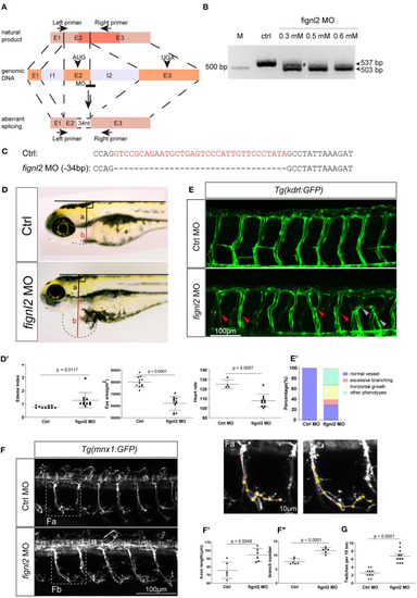
Depletion of fignl2 causes morphological changes in intersegmental vessels (ISVs) and caudal primary neurons. (A) Schematic diagram of morpholino oligonucleotide (MO) induced fignl2 knockdown causing aberrant splicing. (B) MO treatment results in mRNA size change in fignl2 RT-PCR amplicon. The pound sign (#) shows some amplicons of the original size detected after treatment with 0.3 mM MO. (C) MO treatment results in a 34-bp deletion in fignl2 mRNA. (D)fignl2 knockdown causes pericardial edema in developing zebrafish. Graphical representation of edema index (D′), eye size (D′′), heart rate (D′′′) in Ctrl and fignl2 knockdown zebrafish. Ctrl MO, n = 10; fignl2 MO, n = 11. (E)fignl2 knockdown causes morphological changes in ISVs, arrows showing malformations. Graphical representation (E′) of the percentage of different form of ISV malformations caused by fignl2 knockdown. Ctrl MO, n = 10; fignl2 MO, n = 11. (F)fignl2 knockdown causes morphological changes in caudal primary neurons, including greater axon length and increased branching. Representative axons were shown enlarged in (Fa) and (Fb). Graphical representation of axon length (F′), primary branch numbers (F′′) in Ctrl and fignl2 knockdown zebrafish. Ctrl MO, n = 10; fignl2 MO, n = 11. (G)fignl2 mutants shows more frequent twitching. Twitching was counted in 10 s. Ctrl MO, n = 10; fignl2 MO, n = 11.
|
