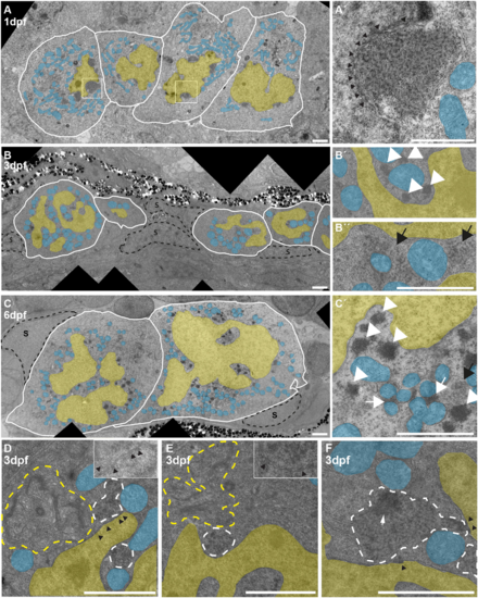
Electron microscopy of wild-type PGCs. (A) PGCs at 1 dpf. Nuclei (yellow) sometimes show one prominent invagination. Nuage can be seen as perinuclear dark patches. (A′) More-detailed view of one nuage patch, showing the granular texture. Black arrowheads indicate nuclear pores, visible as dark stretches within or interrupting the nuclear envelope. (B) PGCs at 3 dpf. The nuclei have acquired an extremely irregular outline and nuage is granular (black arrow, detail in B″) or forms dense granules (white arrowheads, detail in B′) around the nuclei. (C) PGCs at 6 dpf. The nuclei are still heavily gyrated (C′ shows C in more detail). Nuage is mostly very dense, and is found close to the nuclear envelope and in between clusters of mitochondria (white arrowheads and white arrows, respectively). Black arrow indicates granular nuage; S, somatic cells that contact PGCs extensively. (D,E) Two examples of 3 dpf PGCs in which granular nuage (white dashed outline) is in contact with nuclear pores (black arrowheads) and organelles, in this case mitochondria and Golgi (yellow outline). Insets show details without overlays. (F) An example of 3 dpf granular nuage with a more compacted part. Scale bars: 2 µm. Blue overlay indicates mitochondria; yellow overlay indicates nucleus.
|
