
FIGURE
Fig. 1
- ID
- ZDB-FIG-210209-1
- Publication
- Triplett et al., 2020 - Model-based decoupling of evoked and spontaneous neural activity in calcium imaging data
- Other Figures
- All Figure Page
- Back to All Figure Page
Fig. 1
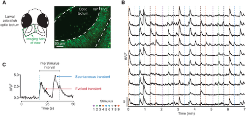
Fig 1. Spontaneous activity in calcium imaging data. (A) Two-photon calcium imaging of the larval zebrafish optic tectum. NP, neuropil; PVL, periventricular layer. (B) Fluorescence traces from 10 example neurons. Dashed vertical lines indicate stimulus onset; colour represents azimuth angle of presented stimulus. (C) Example fluorescence trace segment illustrating that spontaneous calcium transients can occur just before stimulus onset, inflating stimulus-response estimates. |
Expression Data
Expression Detail
Antibody Labeling
Phenotype Data
Phenotype Detail
Acknowledgments
This image is the copyrighted work of the attributed author or publisher, and
ZFIN has permission only to display this image to its users.
Additional permissions should be obtained from the applicable author or publisher of the image.
Full text @ PLoS Comput. Biol.