FIGURE

Figure 1.

ID
ZDB-FIG-210121-1
Publication
Lee et al., 2020 - Suppressive effects of valproic acid on caudal fin regeneration in adult zebrafish
Other Figures
All Figure Page
Back to All Figure Page
Figure 1.

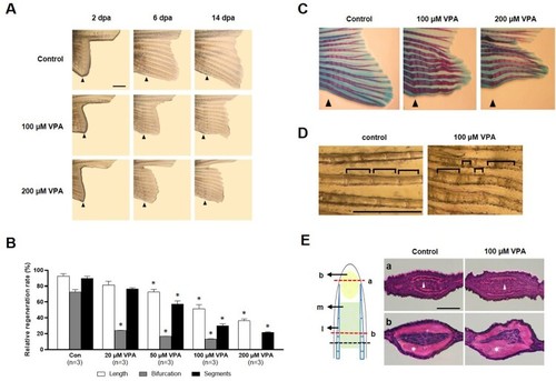
Suppression of caudal fin regeneration after amputation in VPA-treated zebrafish. (A) Images show the regenerated fin in each group at 2, 6, and 14 dpa. Arrowheads indicates the amputation site. Scale bar, 2 mm. (B) Bars indicate the regeneration ratio of length, segments, and bifurcating ray at 14 dpa. Data were expressed as the means ± S.E.M (n = 3). * p < 0.05 compared to the control. (C) Images show Alcian blue and alizarin red staining of cartilages and bones at 14 dpa. (D) Abnormal segments induced by 100 µM VPA treatment during regeneration. Scale bar, 1 mm. (E) Left: The regenerated intra-ray structure is schematically represented. A black dotted line, the amputation site. b, blastemia. m, mesenchyme. l, lepidotrichia. Right: Hematoxylin and eosin staining images show the transverse sections of intra-ray. The irregular-shaped lepidotrichia was shown in 100 µM VPA-treated zebrafish. Asterisks, bony skeletal structure. Arrow heads, actinotrichia. Scale bar, 500 µm.

Expression Data

Expression Detail
Antibody Labeling
Phenotype Data

Phenotype Detail
Acknowledgments
This image is the copyrighted work of the attributed author or publisher, and ZFIN has permission only to display this image to its users. Additional permissions should be obtained from the applicable author or publisher of the image. Full text @ Animal Cells Syst (Seoul)