FIGURE

Figure 2

ID
ZDB-FIG-210108-21
Publication
Hahn et al., 2020 - Expression of the neurotrophic tyrosine kinase receptors, ntrk1 and ntrk2a, precedes expression of other ntrk genes in embryonic zebrafish
Other Figures
All Figure Page
Back to All Figure Page
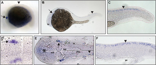
Figure 2

Early expression of <italic>ntrk2a</italic> is seen in the head and spinal cord using whole mount in situ hybridization at 16.5 hpf and 24 hpf.

Expression of ntrk2a is seen in the cranial ganglia (arrows) as well as in the RB neurons of the spinal cord (arrowheads) at (A and D) 16.5 hpf and (B, C, E and F) 24 hpf. The asterisk in E marks weak expression in the diencephalon that matches with previously published accounts. (A, B, C and F) Lateral view. (D and E) Dorsal view. All embryos are oriented with the head to the left. te, marks the telencephalon, mb, marks the midbrain, hb, marks the hindbrain, ov, marks the otic vesicle, chb, marks the caudal hindbrain and ye, marks the yolk extension. Data are representative of two rounds of in situ hybridization with at least 25 embryos per round.

Expression Data
Gene:
Fish:
Anatomical Terms:
Stage Range: 14-19 somites to Prim-5

Expression Detail
Antibody Labeling
Phenotype Data

Phenotype Detail
Acknowledgments
This image is the copyrighted work of the attributed author or publisher, and ZFIN has permission only to display this image to its users. Additional permissions should be obtained from the applicable author or publisher of the image. Full text @ Peer J.