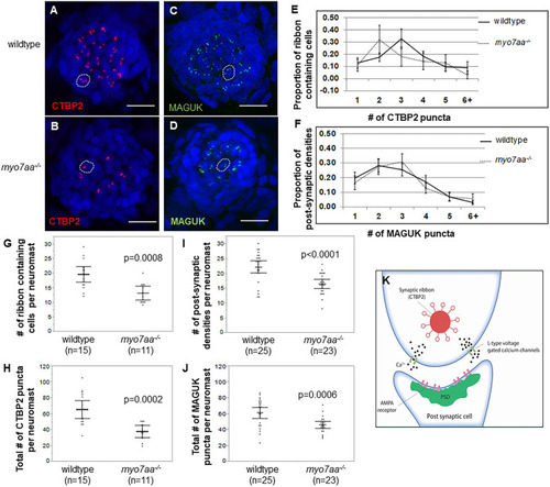
myo7aa−/− Ctbp2 and MAGUK synaptic elements are different fromthose ofwild-typelarvae. (A,B) Representative maximum-intensity projection (z-stack top-down image) of MI1 neuromasts from 5 dpf wild-type larvae (A) and myo7aa−/− larvae (B). Nuclei were stained with DAPI (blue), Ctbp2 is shown in red. Dotted circles outline one hair cell. Scale bars: 10 µm. (C,D) Representative maximum-intensity projection (z-stack top-down image) of MI1 neuromasts from 5 dpf wild type (C) and myo7aa−/− (D). Nuclei were stained with DAPI (blue), MAGUK is shown in green. Dotted circles outline one cell. Scale bars: 10 µm. (E) Distribution of Ctbp2 puncta across a collection of ribbon-containing cells reveals that most 5 dpf wild-type ribbon-containing cells have two Ctbp2 puncta compared to three Ctbp2 puncta in myo7aa−/− hair cells. All error bars are 95% confidence intervals. (F) Distribution of MAGUK puncta across a collection of postsynaptic densities reveals that the proportion of postsynaptic densities is comparable between wild-type and myo7aa−/− larvae. (G,I) 5 dpf wild-type MI1 neuromasts have a greater number of ribbon-containing cells and greater total number of Ctbp2 puncta compared to myo7aa−/− MI1 neuromasts (two-tailed t-test). Bold black lines represent the mean of the data set and error bars are 95% confidence intervals. (H,J) 5 dpf wild-type MI1 neuromasts have a greater number of postsynaptic densities and a greater number of total MAGUK puncta compared to myo7aa−/− MI1 neuromasts (two-tailed t-test). Bold black lines represent the mean of the data set and error bars are 95% confidence intervals. (K) Depiction of the basal end of a hair cell innervated by the auditory nerve. Ctbp2 is alternatively spliced to produce Ribeye protein, the main component of the synaptic ribbon. Ribeye has a halo of tethered vesicles that form the synaptic ribbon (red). Members of the membrane associated guanylate kinase (MAGUK) superfamily are a part of the postsynaptic density (PSD), targeting and anchoring glutamate receptors to the synaptic terminals on the postsynaptic cell (green). Experiments were replicated five times for wild type for both Ctbp2 and MAGUK, and five times for myo7aa−/− mutants for Ctbp2 and two times for MAGUK.
|

