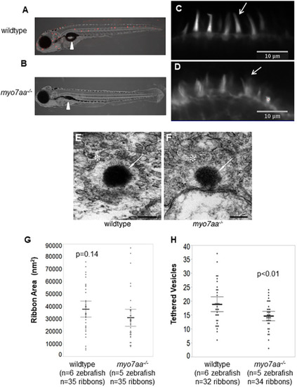
myo7aa−/− larvae have altered mechanotransduction activity, stereocilia structure and ribbon synapse structure. (A,B) Representative light sheet fluorescence microscopy images showing the lateral view of 5 dpf wild-type (A) and myo7aa−/− (B) larvae after brief exposure to FM1-43. FM1-43 staining indicates functional mechanotransduction. Arrowheads point to the swim bladder; myo7aa−/− larvae do not have an inflated swim bladder. (C,D) Representative confocal microscopy images showing the stereocilia of the lateral crista of wild-type (C) and myo7aa−/− (D) larvae at 5 dpf, stained with Alexa-Fluor-488 tagged to phalloidin, a high-affinity filamentous actin probe. Arrows indicate the organized and smooth stereocilia in wild-type hair cells, and disorganized and splayed stereocilia in myo7aa−/− hair cells. Notice that not all stereocilia are splayed in myo7aa−/− hair cells. (E,F) Representative TEM images showing the ribbon synapse structures of 5 dpf wild-type (E) and myo7aa−/− (F) larvae. Arrows point to the ribbon density, stars indicate a halo of tethered vesicles in the inner ear hair cells. Scale bars: 200 nm. (G) Wild-type ribbon synapses have a comparable ribbon area to myo7aa−/− ribbon synapses (two-tailed t-test). (H) Wild-type ribbon synapses have an increased number of tethered vesicles compared to myo7aa−/− ribbon synapses (two-tailed t-test). The bold black lines represent the mean of the data set and error bars are 95% confidence intervals. Electron microscopy experiments were replicated four times for wild type and three times for myo7aa−/− mutants.
|
