
Figure 9
- ID
- ZDB-FIG-200814-40
- Publication
- Lee et al., 2020 - Methiothepin Suppresses Human Ovarian Cancer Cell Growth by Repressing Mitochondrion-Mediated Metabolism and Inhibiting Angiogenesis In Vivo
- Other Figures
- All Figure Page
- Back to All Figure Page
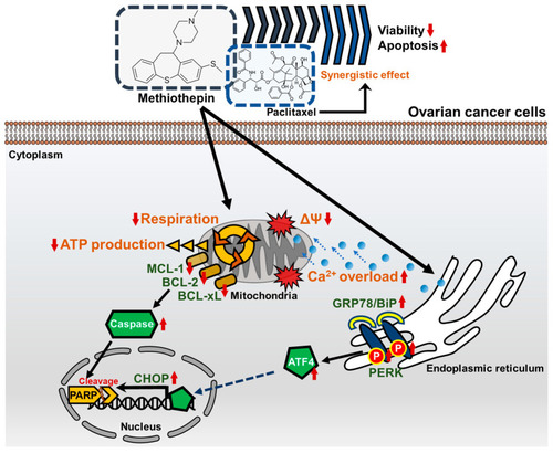
Schematic diagram of the methiothepin-associated anti-cancer mechanisms in ovarian cancer cells. In ovarian cancer cells, methiothepin induces depolarization in the mitochondrial membrane (also known as mitochondria membrane potential, ΔΨ) and promotes the influx of Ca2+. Metabolic disorders caused by methiothepin are indicated by a decrease in mitochondrial respiration and adenosine triphosphate (ATP) production. The expression of proteins belonging to the BCL-2 family are inhibited by methiothepin, which leads to the cleavage of poly (ADP-ribose) polymerase (PARP), along with the activation of caspases. Meanwhile, methiothepin increases the expression of ER stress-related proteins and finally induces the expression of C/EBP homologous protein (CHOP), which leads to apoptosis. Methiothepin improves the anti-cancer effect of paclitaxel, greatly reducing the viability of ovarian cancer cells. |