FIGURE

Figure 2

ID
ZDB-FIG-200814-34
Publication
Lee et al., 2020 - Methiothepin Suppresses Human Ovarian Cancer Cell Growth by Repressing Mitochondrion-Mediated Metabolism and Inhibiting Angiogenesis In Vivo
Other Figures
All Figure Page
Back to All Figure Page
Figure 2

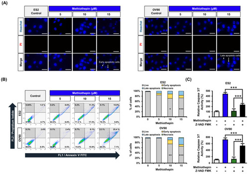
Regulation of cell death by methiothepin in ES2 and OV90 cells. (A) Hoechst (blue) and PI (red) staining was performed for cell death analysis in ES2 and OV90 cells. Scale bar indicates 40 μm. (B) Annexin V and PI staining were used to measure the death of ES2 and OV90 cells following methiothepin treatment. The percentage of cell distribution observed by flow cytometry was determined for the apoptotic pattern in each quadrant. The lower left quadrant corresponds to live cells, the upper left, to necrosis, the lower right, to early apoptosis, and the upper right, to late apoptosis. (C) The activity of caspase 3/7 in ES2 and OV90 cells treated with methiothepin was measured following the treatment of the cells with the pan-caspase inhibitor Z-VAD FMK. Results are expressed as the means ± SDs of three independent experiments. The asterisks indicate statistically significant differences compared to the control (* p < 0.05; ** p < 0.01; *** p < 0.001).

Expression Data

Expression Detail
Antibody Labeling
Phenotype Data

Phenotype Detail
Acknowledgments
This image is the copyrighted work of the attributed author or publisher, and ZFIN has permission only to display this image to its users. Additional permissions should be obtained from the applicable author or publisher of the image. Full text @ Pharmaceutics