
Figure 7
- ID
- ZDB-FIG-200814-25
- Publication
- Lee et al., 2020 - Methiothepin Suppresses Human Ovarian Cancer Cell Growth by Repressing Mitochondrion-Mediated Metabolism and Inhibiting Angiogenesis In Vivo
- Other Figures
- All Figure Page
- Back to All Figure Page
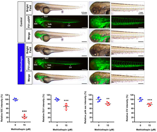
Impairment of vasculogenesis by methiothepin exposure in zebrafish embryos. Fluorescence images of transgenic (fli1:eGFP) zebrafish embryos after treatment with methiothepin for 48 h were captured by fluorescent microscopy. Quantification of fluorescence intensity in the vascular structures of the inner optic circle (IOC), dorsal longitudinal vein (DLV), yolk sac, and caudal vein (CV) of zebrafish embryos following methiothepin exposure was performed using ImageJ software. The asterisks indicate statistically significant differences compared to the control (** |