
FIGURE
Fig. 4
- ID
- ZDB-FIG-200707-3
- Publication
- Nadar et al., 2020 - Preclinical evaluation of platinum-loaded hydroxyapatite nanoparticles in an embryonic zebrafish xenograft model
- Other Figures
- All Figure Page
- Back to All Figure Page
Fig. 4
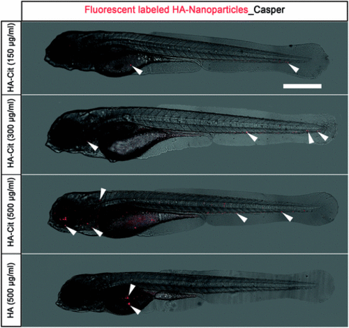
Biodistribution of fluorescently labeled HA-Cit and HA nanoparticles in zebrafish embryo. Biodistribution of fluorescently labeled HA-nanoparticles (in red) injected in casper embryos at 2 days post injection (2 dpi). At concentrations between 150–500 μg ml−1, all citrate-functionalized HA-Cit nanoparticles (top) were spread homogeneously throughout the embryos, whereas citrate-free HA nanoparticles accumulated near the injection site (bottom). HA nanoparticles are depicted in red, white arrowheads correspond to fluorescently labeled HA nanoparticles. Scale bar: 500 μm. |
Expression Data
Expression Detail
Antibody Labeling
Phenotype Data
Phenotype Detail
Acknowledgments
This image is the copyrighted work of the attributed author or publisher, and
ZFIN has permission only to display this image to its users.
Additional permissions should be obtained from the applicable author or publisher of the image.
Full text @ Nanoscale