
Fig. 3
- ID
- ZDB-FIG-200707-2
- Publication
- Nadar et al., 2020 - Preclinical evaluation of platinum-loaded hydroxyapatite nanoparticles in an embryonic zebrafish xenograft model
- Other Figures
- All Figure Page
- Back to All Figure Page
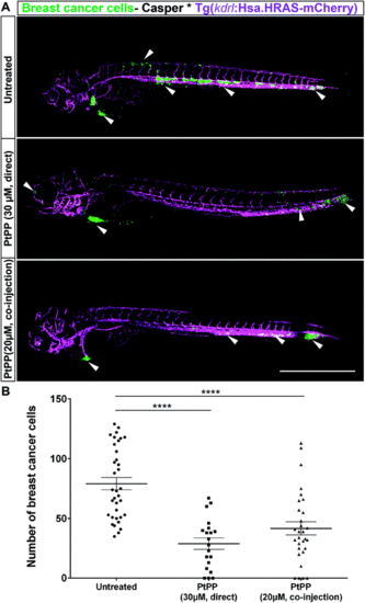
Effect of direct addition or co-injection of PtPP on zebrafish embryo hosting breast cancer cells. (A) Representative images of zebrafish embryo expressing vascular marker Tg(kdrl:Has.HRAS-mCherry) in casper background hosting eGFP labeled breast cancer cells (MDA-MB-231_eGFP) at 2 days post injection: in contrast to untreated Pt-free controls, direct addition of 30 μM PtPP (center) and co-injection of 20 μM PtPP (bottom) revealed a reduction in breast cancer cell number. Vasculature is indicated in magenta and breast cancer cells are depicted in green. White arrowheads correspond to presence of cancer cells in the respective regions. Scale bar: 500 μm. (B) Manual quantification of cancer cells at 2 days post injection revealing a significant reduction of breast cancer cell number in PtPP-treated embryos compared to controls. Plot represents mean ± SEM. Statistical analysis: one-way ANOVA followed by Dunnett's test for multiple comparisons. ****P < 0.0001. |