|
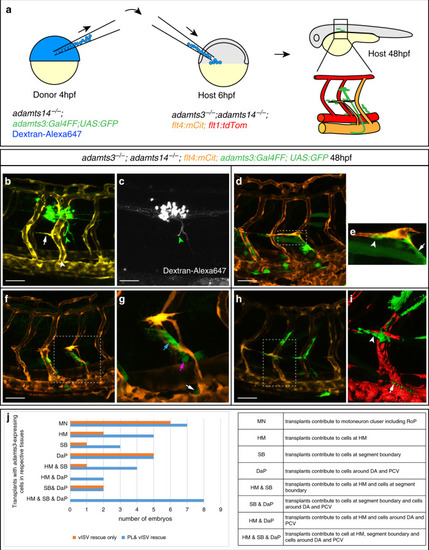
<italic>adamts3</italic> expression via motoneurons or mesenchymal cells can rescue venous sprouting and PL formation in <italic>adamts3</italic>; <italic>adamts14</italic> double mutants.a Diagram depicting the heterochronic transplantation approach: Dextran-Alexa647 is injected into one-cell stage adamts14−/−; adamts3:Gal4FF; UAS:GFP embryos (donor embryos). Cells are transferred from donor embryos to adamts3; adamts14 double mutants at 6 hpf (host embryo) and the effects are assessed at 48 hpf. b, c In cases where transplanted cells contributed to adamts3-expressing motoneuron clusters (green arrowhead), venous ISVs (white arrowhead), and PL cells (white arrow) formed in double mutants. c Transplanted cells labeled by Dextran-Alexa647. d When transplants gave rise to adamts3+ cells at three locations, the HM, the ventral segment boundaries, and close to the DA and PCV, a local rescue of the formation of PL cells and intersegmental veins in adamts3; adamts14 double mutant embryos was visible. e Zoom-in of the boxed region in (d) with adamts3-expressing cells at the segment boundary highlighted by an arrow and transplanted cells at the HM marked by an arrowhead. f, g Transplantation of cells contributing to adamts3-expressing cells at the ventral segment boundaries (blue arrow) and to cells located close to the DA (magenta arrow) and the PCV (white arrow) also resulted in a locally restricted rescue of PL and vISV formation. g Zoom-in of the indicated region in f. h, i Cases, in which the transplants gave rise to adamts3+ cells at the HM (arrowhead in i) and the region around the PCV (arrow in i), but not to the cells along the ventral segment boundaries showed a rescue of PL cells and vISVs. j Overview about the results obtained from 23 rounds of cell transplantations. Each round contained 144 recipient embryos obtained from an adamts3+/−; adamts14−/− incross of which a quarter was expected to be double mutant (in total 828). Embryos showing a rescue of PL or vISV development were selected for imaging and genotyping. All embryos shown in (j) are adamts3; adamts14 double mutants. Scale bars: 50 µm. DA dorsal aorta, hpf hours post fertilization, PCV posterior cardinal vein, PL parachordal lymphangioblast, vISV venous intersegmental vessel.
|