|
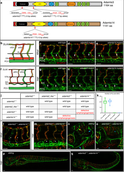
Lymphangiogenesis is completely abolished in <italic>adamts3</italic>; <italic>adamts14</italic> double mutant embryos.a Schematic of Adamts3 and Adamts14 protein structures depicting the predicted effects of the indicated deletion alleles. Shown are the aa positions of the deletion-induced frame shifts (red letters) as well as the position of the resulting premature stop codons. SP signal peptide, MPD metallopeptidase domain, ACR ADAM cysteine-rich domain 2, TSP1 thrombospondin type-1. b Schematic representation of the wild-type trunk vasculature at 48 hpf. c–e, g–i, and l–qflt4:mCitrine; flt1:tdTomato double transgenic embryos highlighting arterial ECs in red and venous and lymphatic structures in green. In wild-type (c), adamts3 (d) or adamts14 single mutants (e), PL cells align at the HM at 48 hpf (arrows). f Schematic representation of the trunk vasculature at 5 dpf with the thoracic duct (TD) being located between DA and PCV. Compared with wild type (g), neither homozygous adamts3 (h) nor adamts14 mutants (i) exhibit lymphatic defects at 5 dpf (arrows). j Table summarizing the analysis of lymphatic phenotypes in all double mutant combinations for adamts2, adamts2_like, adamts3, and adamts14. k At 48 hpf, the number of PLs was quantified in n = 53 siblings and n = 30 adamts3; adamts14 double mutants and the number of vISVs was quantified in n = 37 siblings and n = 11 double mutants. Box-and-whisker plots show median and quartiles with whiskers indicating minimum/maximum values (source data are provided as a Source Data file). ladamts3; adamts14 double mutants lack PLs (white asterisks) and do not form vISVs (blue asterisks) at 48 hpf. m At 5 dpf, the TD is completely absent in double mutants (white asterisks). Facial lymphatics (blue arrows) and meningeal lymphatics (yellow arrow) are formed in siblings at 5 dpf (n) but are absent in adamts3; adamts14 double mutants (o). In contrast to their siblings (p), double mutants (q) display a variably strong curvature of the trunk. Scale bars in c–e, g–i, l–o: 50 µm; p, q: 100 µm. aa amino acid, bp base pair, dpf days post fertilization, ECs endothelial cells, DA dorsal aorta, DLAV dorsal longitudinal anastomotic vessel, hpf hours post fertilization, HM horizontal myoseptum, a/vISV arterial/venous intersegmental vessel, PCV posterior cardinal vein, PL parachordal lymphangioblast, VTA vertebral artery.
|