|
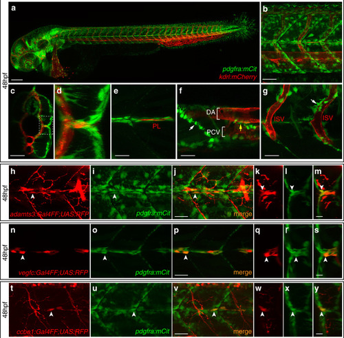
Co-expression of <italic>adamts3</italic>, <italic>ccbe1</italic>, and <italic>vegfc</italic> with <italic>pdgfra</italic> at the horizontal myoseptum.a–g Confocal images of pdgfra:mCitrine; kdrl:mCherry double transgenic embryos at 48 hpf. a Lateral view giving an overview of the pdgfra-expression domains. b Higher magnification of the trunk region above the yolk extension. c, d Virtual cross section of the trunk shown in b. d Zoom-in of the boxed area in (c) showing the HM region in detail. e–g Partial projections of (b). e At the HM, pdgfra-expressing cells (in green) are surrounding the PL cells (in red). fpdgfra is expressed by cells located along the segment boundaries (white arrow) as well as by individual cells located around the main axial blood vessels (yellow arrow). g At the level of the ISVs, individual pdgfra+ cells locate in close proximity to the blood vessels. h–j, n–p, and t–v Lateral views and k–m, q–s, and w–y virtual cross sections of the HM region in double transgenic embryos at 48 hpf. White arrows highlight cells with co-expression. h–m A subpopulation of pdgfra-expressing cells (green) is co-labeled by the adamts3 reporter transgene (red) at the HM. n–s Cells at the HM co-express vegfc (red) and pdgfra (green). t–y All ccbe1-expressing cells at the HM (red) show co-expression of pdgfra (green). Scale bar in a: 100 µm; b, c: 50 µm; e–j, n–p, t–v: 25 µm; k–m, q–s, w–y: 10 µm. DA dorsal aorta, HM horizontal myoseptum, hpf hours post fertilization, ISV intersegmental vessel, PCV posterior cardinal vein, PL parachordal lymphangioblast.
|