FIGURE

Figure 3—figure supplement 2.

ID
ZDB-FIG-200530-6
Publication
Williams et al., 2020 - Nodal and Planar Cell Polarity signaling cooperate to regulate zebrafish convergence and extension gastrulation movements
Other Figures
All Figure Page
Back to All Figure Page
Figure 3—figure supplement 2.

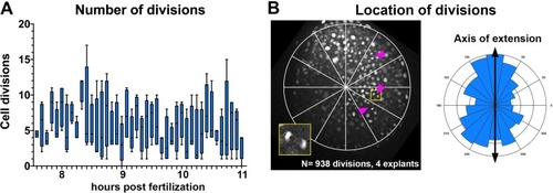
Cell divisions within <italic>ndr2</italic>-expressing explants.

(A) Manually annotated cell divisions (per explant) within four live explants from WT embryos co-injected with H2B-RFP and 10 pg ndr2 RNA. Measurements were taken at 5 min intervals from 7.5 to 11 hpf. Boxes represent the 25th to 75th percentiles, whiskers are minimum and maximum vales, bars are median values. (B) Left: representative image of cell divisions (arrowheads) within an explant co-expressing H2B-RFP and ndr2. The inset is enlarged from the region in the yellow square. Right: radial histogram of locations of all cell divisions (with respect to the center of each explant) detected within four ndr2-expressing explants between 7.5 and 11 hpf, with 90°/270° representing the axis of extension.

Expression Data

Expression Detail
Antibody Labeling
Phenotype Data

Phenotype Detail
Acknowledgments
This image is the copyrighted work of the attributed author or publisher, and ZFIN has permission only to display this image to its users. Additional permissions should be obtained from the applicable author or publisher of the image. Full text @ Elife