|
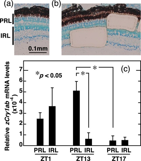
qRT-PCR analysis of zCry1ab mRNA levels in the zebrafish retinal cells captured by laser microdissection (LMD). Retinas were collected at ZT1 or ZT13 or ZT17 from zebrafish entrained to long-day cycles (14L10D). Photoreceptor cell layer (PRL) mainly containing outer nuclear layer (visual photoreceptors) and inner retinal layer (IRL) containing inner nuclear layer, inner plexiform layer and retinal ganglion cells were obtained from the retinal sections. (a) A retinal section (ZT1) showing PRL and IRL. (b) An example of retinal sections after the collection of PRL and IRL samples by LMD. (c) Levels of zCry1ab mRNA in PRL and IRL. The levels of zCry1ab mRNA and 18S rRNA were measured by qRT-PCR, and the zCry1ab mRNA levels relative to 18S rRNA levels were shown. Error bars represent ± SE. Data were analyzed by two-way ANOVA (Supplementary Table S72) and Tukey-Kramer post-hoc tests (Supplementary Table S73). Asterisks represent significant difference (p < 0.05).
|