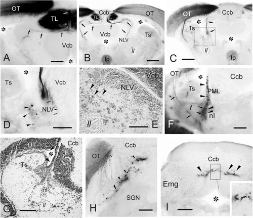
Labeled cells and fibers after direct DiI application to TL. Photomicrographs of transverse sections caudal to the tracer application point showing labeled structures observed after homolateral DiI application to intermediate TL. Ipsilateral side is to the left. (A–C) Sections showing the caudal course of the torovalvular tract (arrows) from the labeled TL (A,B) that enters and travels bilaterally through the cerebellar valvula. Note in (C) the (tecto-)isthmal tract (white arrow) also labeled from some DiI diffusion to the ipsilateral optic tectum. (D) Detailed view showing labeled cells ventrolaterally to the nucleus lateralis valvulae (arrowheads) squared in (C), which could represent the subvalvular nucleus in the zebrafish. Note a few labeled fibers from the torovalvular tract traversing the nucleus lateralis valvulae (arrows). (E) Nissl stained section showing larger cell bodies (putative subvalvular nucleus, arrowheads) ventrolaterally to the nucleus lateralis valvulae. (F) Labeled cells (arrowheads) and fibers (arrows) in the posterior mesencephalic lamina and the nucleus isthmi. (G) Nissl stained section showing the posterior mesencephalic lamina (arrowheads). (H) Section showing labeled cells and fibers in the optic tectum, as well as cells in the cerebellar body (arrowheads). (I) Labeled cells in the granule cell layer of the cerebellar body (arrowheads), detailed in the inset. Asterisk: ventricle. For abbreviations, see list. Scale bars: 200 μm (B,C,F–I); 100 μm (A,D,E).
|
