|
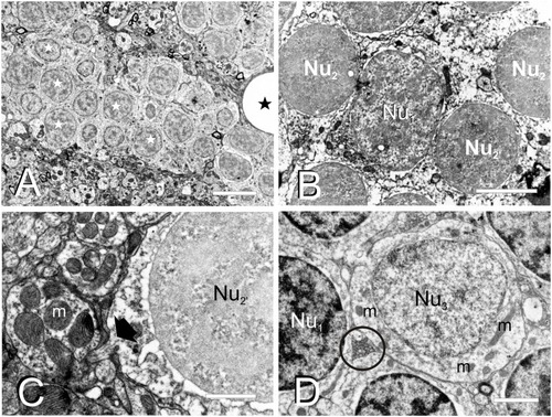
Fine cell structure of the adult TL. (A–D) Electron micrographs showing the main three nucleus types found in TL. (A) Low magnification electron micrograph showing a cell cluster with medium-sized round nuclei with partially condensed chromatin (Nu1, white star) and surrounded by a dense neuropil. (B) Detail of a medium-sized nucleus (Nu1) surrounded by smaller nuclei (Nu2 and Nu2’). (C) Detail of a small-sized cell nucleus with non-homogeneously condensed chromatin (Nu2’) showing the exit of the axon (black arrow) from the cell body. (D) Nucleus (Nu3) with pale chromatin that belongs to the large-sized cells among other nuclei with more condensed chromatin (Nu1). Note also the small compact bundle of non-myelinated axons (circle). m, mitochondria; black stars, blood vessels. Scale bars: 5 μm (A), 2 μm (B,D), 1 μm (C).
|