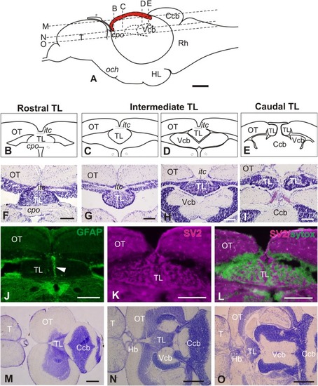
General morphology of the adult zebrafish Torus Longitudinalis (TL). (A) Schema of the brain in a lateral view representing the location of the TL (in red) along the rostro-caudal axis. Anterior to the left. The approximate levels of sections (B–D) and (K–M) are also shown. (B–E) Diagrams of the general anatomy of the TL at rostral, intermediate and caudal levels in transverse section. (F–I) Images of Nissl stained sections showing TL at approximately the same rostrocaudal levels as (B–E). (J) Glial processes along TL midline (arrowhead) after GFP immunostaining in Tg(gfap:GFP)mi2001. (K) Neuropil in TL showing immunoreactivity against synaptic vesicles 2 (SV2). (L) Merged channels for SV2 (magenta) and Sytox green (green). Note that the non-immunoreactive areas in (I) correspond with location of cell bodies in green). (M–O) Horizontal sections [at levels indicated in (A)] showing the general anatomy and location of TL. Anterior to the left. For abbreviations, see list. Scale bars: 100 μm (A-L), 250 μm (M-O).
|
