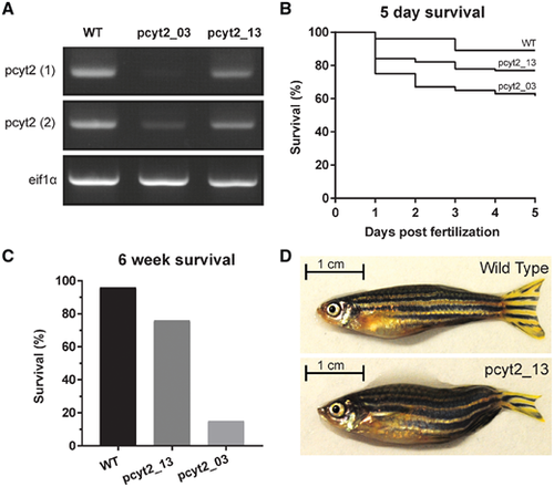
CRISPR zebrafish PCYT2 model. Two transgenic zebrafish lines were created by knocking out the third (pcyt2_03) and final 13th exon (pcyt2_13), respectively. (A) Complementary DNA expression analysis of pcyt2 using two primer sets in wild-type (WT), pcyt2_03 and pcyt2_13 showing low/absent pcyt2 expression in pcyt2_03 and moderately reduced pcyt2 expression in pcyt2_13. eif1α was used as loading control. (B) Kaplan-Meier plot for the survival of the first 5 days post-fertilization, significance was calculated using the log rank and Wilcoxon test [χ2for equivalence of death rates: wild-type versus pcyt2_03 = 21.440258 (P < 0.0001), wild-type versus pcyt2_13 = 6.497151 (P = 0.0108) and pcyt2_03 versus pcyt2_13 = 4.700852 (P = 0.0301)]. (C) Survival at 6 weeks post-fertilization for wild-type, exon 3 and exon 13 deletion showing a significantly higher survival for the exon 13 deletion mutant, significance calculated via fisher exact test (P < 0.001). (D) The pcyt2_13 line compared with the wild-type at 6 weeks of age showing smaller overall size and abnormal tail-fin morphology.
|
