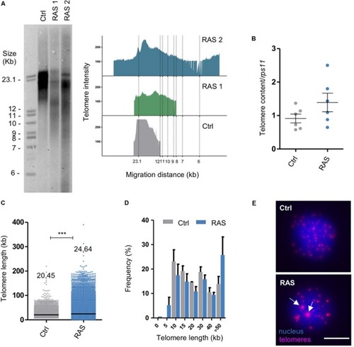
Zebrafish brain tumors have heterogeneous telomeres. (A) Telomere length analysis via TRF in one control and two RAS tumors. The panel on the right shows TFR analysis obtained by graphing intensity of the signal versus DNA migration. (B) Relative telomere content determined by telomere qPCR and normalized to the signal of a single copy gene (rps11) in controls (Ctrl, n = 7 brains) and RAS tumors (RAS, n = 7 brains). Bars represent mean ± SEM. (C) Quantification of relative telomere length measured by Q-FISH and given a kb value based on the fluorescent intensity of L5178Y-S and L5178Y-R lymphocyte cells with known telomere lengths of 10.2 and 79.7 kb, respectively. See also Supplementary Figure 1B. Number of telomeres examined: Ctrl n = 3027, RAS = 9738. Data from three independent experiments were combined. Median values are reported on the graph. ***p < 0.0001. Scatterplot bars represent median. (D) Distribution of telomere length evaluated by Q-FISH in Control (gray n = 2) and RAS tumors (light blue n = 3). The very long telomeres (>30 kb) could represent telomeric clusters, an ALT feature. (E) Representative fluorescent microscope images of Q-FISH analysis of control and RAS nuclei with ultrabright foci (white arrows). Scale bar: 5 μm. TRF: telomere restriction fragment; Ctrl: control; RAS: brain tumors. See also Supplementary Figure 1.
|
