
Fig. 5
- ID
- ZDB-FIG-200302-58
- Publication
- Librán-Pérez et al., 2019 - Antiviral activity of palmitic acid via autophagic flux inhibition in zebrafish (Danio rerio)
- Other Figures
- All Figure Page
- Back to All Figure Page
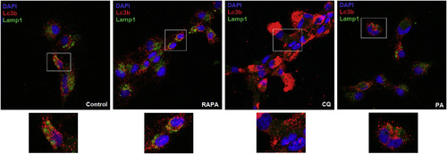
Palmitic acid inhibits autophagosome-lysosome fusion. The ZF4 cells under conditions of starvation were incubated with PA, RAPA or CQ for 2.5 h. Untreated controls were also included. After the incubation period, the cells were fixed and immuno-stained for Lc3b (red) and Lamp1 (green) detection. Nuclei were stained with DAPI (blue). Images were taken by confocal microscopy to determine the prevalence of the co-localization of Lc3b and Lamp1. Representative images were selected to reflect the effect of the different compounds on the autophagic flux. Yellow/orange puncta indicate co-localization of Lc3b and Lamp1 (autophagic flux). (For interpretation of the references to colour in this figure legend, the reader is referred to the Web version of this article.) |