FIGURE

Fig. 3

ID
ZDB-FIG-200213-13
Publication
Shibata-Germanos et al., 2019 - Structural and functional conservation of non-lumenized lymphatic endothelial cells in the mammalian leptomeninges
Other Figures
All Figure Page
Back to All Figure Page
Fig. 3

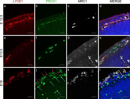
Cells expressing LLEC markers are present within embryonic mouse leptomeninges. ad As revealed by IHC, E12.5 mouse meninges (white bracket) contain cells that co-express LYVE1 (red), PROX1 (green), and MRC1 (white). DAPI marks the nuclei in blue (d). Scale = 30 µm. n = 3 brains. eh E13.5 mouse meninges (white bracket) contain cells that co-express LYVE1 (red), PROX1 (green), and MRC1 (white). DAPI marks the nuclei in blue (h). Scale = 30 µm. n = 3 brains. il E18 mouse meninges (white bracket) contain cells that co-express LYVE1 (red), PROX1 (green), and MRC1 (white). PROX1 is cytoplasmic (j, red arrows) in the meninges but nuclear in neurons (j, blue arrows). DAPI marks the nuclei in blue (l). Scale = 30 µm. n = 3 brains

Expression Data

Expression Detail
Antibody Labeling
Phenotype Data

Phenotype Detail
Acknowledgments
This image is the copyrighted work of the attributed author or publisher, and ZFIN has permission only to display this image to its users. Additional permissions should be obtained from the applicable author or publisher of the image. Full text @ Acta Neuropathol.