|
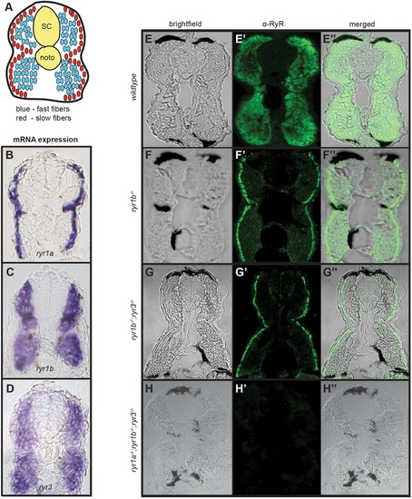
Mutant ryr alleles do not produce protein products. (A) Schematic of a cross-section of the trunk of a 1-2 dpf zebrafish embryo highlighting muscle organization. SC spinal cord, noto notochord. (B-D) Transverse cryosections of 24 hpf embryo trunks illustrating the RNA expression patterns of (B) ryr1a, (C) ryr1b, and (D) ryr3 detected by WISH. (E-H″) Transverse sections through the trunks of 48 hpf wild-type and mutant embryos immunostained with the 34C (anti-RyR) antibody. Brightfield images (E-H), fluorescent images showing 34C staining (green) (E′-H′), and merged brightfield/fluorescent images (E″-H″) of embryos of indicated genotypes. Each ryr mutation is associated with loss of a distinct component of the normal expression pattern of RyR channels in embryonic muscle. Somitic muscle of triple ryr1a;ryr1b;ryr3 mutants lack all RyR protein detected by the 34C antibody.
|