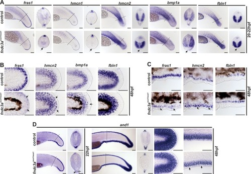
Normal development of the ventral median fin fold is altered in fndc3awue1/wue1 mutants. (A) Investigation of fras1, hmcn1, hmcn2, bmp1a, and fbln1 expression in the tail bud 20–22 hpf indicated loss of mesenchymal cells in the median fin fold in fndc3awue1/wue1 mutants (arrows; fras1: 9/32; hmcn1: 11/26; hmcn2: 24/43; bmp1a: 22/45; fbln1: 16/32). (B) Expression of mesenchymal markers in posterior fins of 48 hpf old embryos was slightly changed following loss of fndc3a (fras1: 10/24; hmcn2: 12/25; bmp1a: 8/21; fbln1: 9/24). (C) Ventral fin folds in 48 hpf old embryos did not show loss of mesenchymal markers after fndc3awue1/wue1 mutation. (D) Expression of and1, an essential marker for actinotrichia formation, in fndc3awue1/wue1 in the ventral fin fold was partly lost in 22 hpf embryos (12/25), but recovered in 48 hpf old embryos to a reduced expression level (control n = 16; weaker expression in fndc3awue1/wue1 embryos n = 10/18). Dashed lines in (A,D) indicate planes of shown sections. Dashed lines in (B,C) indicate fin boundaries. Scale bars: 50 µm
|
