
Figure 5
- ID
- ZDB-FIG-191230-1294
- Publication
- Suen et al., 2019 - IL-10 from plasmacytoid dendritic cells promotes angiogenesis in the early stage of endometriosis
- Other Figures
- All Figure Page
- Back to All Figure Page
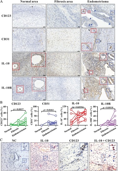
Analysis of CD123, CD31, IL‐10, and IL‐10R expression in human endometrioma and uterine endometrial tissues. (A) Immunohistochemistry for CD123, CD31, IL‐10, or IL‐10R (brown colour) in a representative endometrioma lesion as well as the fibrosis area and normal area surrounding the corresponding endometrioma lesion. The insets (solid box) show two‐fold enlarged images of vascular structure (V) or glandular epithelium (G) (dashed box) with the same colour. Arrows indicate CD123+ cells. Blue, haematoxylin counterstain. Black scale bars = 50 µm. The enlarged images of IL‐10 and IL‐10R expression on endometrioma lesions in A are shown in supplementary material, Figure |