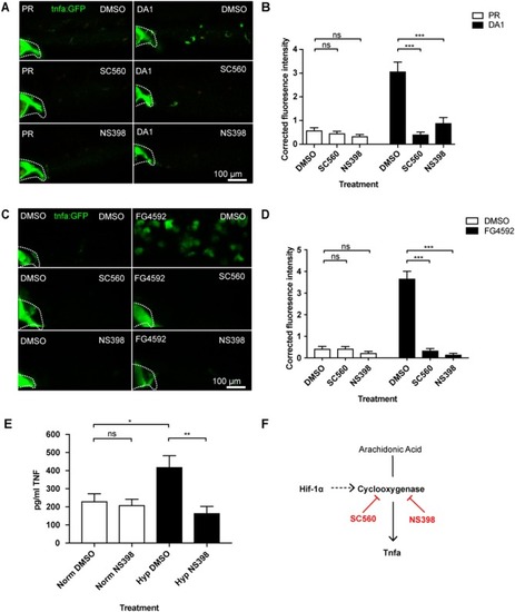
Hif-1α-activated tnfa is cyclooxygenase dependent. (A) Confocal micrographs of 2 dpf caudal vein region of larvae in the TgBAC(tnfa:GFP)pd1028 line. Phenol red (PR) and dominant active Hif-1α (DA1) injected larvae were treated with DMSO, SC560 (Cox-1 inhibitor), and NS398 (Cox-2 inhibitor). Dotted lines indicate the yolk extension of the larvae where there is non-specific fluorescence. (B) Corrected fluorescence intensity levels of tnfa:GFP in larvae in (A). Mean ± SEM, n = 36 cells from six embryos representative of three independent experiments. P-values shown are: *P < 0.05, **P < 0.01, and ***P < 0.001, two way ANOVA with Bonferonni post-test adjustment. (C) Confocal micrographs of 2 dpf caudal vein region of larvae in the TgBAC(tnfa:GFP)pd1028 line. DMSO and FG4592 treated larvae were co-treated with DMSO, SC560 (Cox-1 inhibitor), and NS398 (Cox-2 inhibitor). Dotted lines indicate the yolk extension of the larvae where there is non-specific fluorescence. (D) Corrected fluorescence intensity levels of tnfa:GFP in larvae in (C). Mean ± SEM, n = 48 cells from eight embryos representative of three independent experiments. P-values shown are: *P < 0.05, **P < 0.01, and ***P < 0.001, two way ANOVA with Bonferonni post-test adjustment. (E) TNF ELISA of human monocyte derived macrophages treated with LPS and incubated in normoxia or 0.8% hypoxia with or without treatment with NS398. LPS negative controls were performed but TNF produced in these groups was below detectable levels. Mean ± SEM, n = 5–8 biological repeats from 3 to 4 donors. P-values shown are: *P < 0.05, **P < 0.01, and ***P < 0.001, two way ANOVA with Bonferonni post-test adjustment. (F) Schematic of the arachidonic pathway indicating that stabilizing Hif-1α upregulates tnfa, an effect that is blocked using the Cox1/2 inhibitors SC560/NS398.
|
