
FIGURE
Fig. S1
- ID
- ZDB-FIG-191025-1
- Publication
- Liverani et al., 2017 - Innovative approaches to establish and characterize primary cultures: an ex vivo 3D system and the zebrafish model.
- Other Figures
- All Figure Page
- Back to All Figure Page
Fig. S1
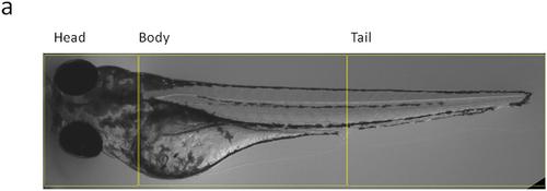
Brightfield image of a zebrafish embryo, showing an example of segmentation into 3 different anatomical regions (head, body and tail) in which the engrafted foci of the injected patient-derived cells were quantified. |
Expression Data
Expression Detail
Antibody Labeling
Phenotype Data
Phenotype Detail
Acknowledgments
This image is the copyrighted work of the attributed author or publisher, and
ZFIN has permission only to display this image to its users.
Additional permissions should be obtained from the applicable author or publisher of the image.
Full text @ Biol. Open