|
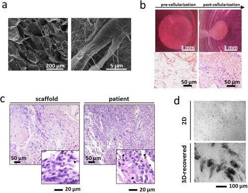
Characterization of the ex vivo 3D tumor model. (A) Scanning electron microscopy (SEM) analysis of collagen-based scaffolds at different magnifications. Images were taken with a FEI Nova NanoSEM microscope. (B) Pictures of collagen scaffolds pre- and post-cellularization with primary liposarcoma cells and hematoxylin & eosin staining of paraffin-embedded sections of the scaffold pre- and post-cellularization (C) Hematoxylin & eosin staining of paraffin-embedded sections of 3D scaffolds cultured with primary liposarcoma cells and of the histological specimen. Arrowheads indicate tumor cells. (D) Inverted microscopy pictures of 2D-cultured and 3D-recovered liposarcoma cells.
|