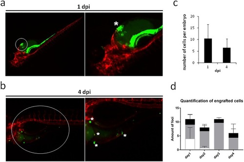
Representative stereo micrograph images of primary liposarcoma cells (green, CFSE) injected into 2 dpf Tg(Kdrl:mCherry) zebrafish embryos. Images taken at (A) 1 dpi and (B) 4 dpi. White circles indicate the area zoomed in the close-ups; white asterisks indicate the injected cells (A) and the invading cells (B). The number of engrafted foci per embryo is reported. Quantification of total number of engrafted foci at 1 and 4 dpi (C) (mean±s.d., n=4) and quantification of engrafted foci in the three different anatomical regions of the zebrafish embryos (D) (mean±s.d., n=3). White, tail region; gray, body region; black, head region. At 4 dpi, we detected that liposarcoma-derived cells survived in vivo and spread from the injection site. Of note, injected embryos showed an aspecific CFSE signal in the gastrointestinal trait of the embryos, at the initial timepoint of the experiment (1 dpi), due to dye leakage. This aspecific signal faded out at the later timepoint, as visible in the pictures of 4 dpi embryos.
|
