
Fig. 1
- ID
- ZDB-FIG-191010-14
- Publication
- Petrachkova et al., 2019 - Lack of Cyclin B1 in zebrafish causes lengthening of G2 and M phases
- Other Figures
- All Figure Page
- Back to All Figure Page
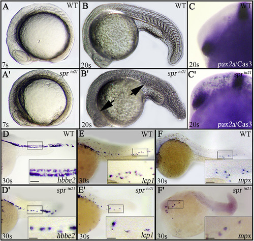
The spr mutant phenotype. (A, B) Morphology of the live embryo, side view of: (A, B) wild-type and (A′, B′) sprtu21 embryos. Arrows indicate darkening in the head and trunk. Note the undefined somiteborders in the mutant by 20-somites (19 h). (C, C′) Apoptosis, visualized by antibody staining for an active form of Caspase 3, is evident in the head of the spr mutant by 25-somites (21.5 h). Embryos are shown in side view using pax2a mRNA expression to define the area between the hindbrain and optic stalk. (D-F′) Blood cells are larger and less frequent in the spr mutant. (D, D′) Labeling of the blood by the erythrocyte marker, hbbe2 (Brownlie et al., 1998), 8/8 mutants and 20/20 wild-type embryos displayed this phenotype; (E, E′) the macrophage marker, lcp1 (Herbomel et al., 1999), 10/10 mutants and 17/17 wild-type embryos displayed this phenotype; (F, F′); and the neutrophil marker, mpx (Bennett et al., 2001; Lieschke et al., 2001), 10/10 mutants and 21/21 wild-type embryos displayed this phenotype. Boxes indicate the areas enlarged at higher magnification, scale bar is 20 μm. |
| Genes: | |
|---|---|
| Fish: | |
| Anatomical Terms: | |
| Stage: | Prim-5 |
| Fish: | |
|---|---|
| Observed In: | |
| Stage Range: | 5-9 somites to Prim-5 |
Reprinted from Developmental Biology, 451(2), Petrachkova, T., Wortinger, L.A., Bard, A.J., Singh, J., Warga, R.M., Kane, D.A., Lack of Cyclin B1 in zebrafish causes lengthening of G2 and M phases, 167-179, Copyright (2019) with permission from Elsevier. Full text @ Dev. Biol.