
Fig. 7
- ID
- ZDB-FIG-191010-9
- Publication
- Petrachkova et al., 2019 - Lack of Cyclin B1 in zebrafish causes lengthening of G2 and M phases
- Other Figures
- All Figure Page
- Back to All Figure Page
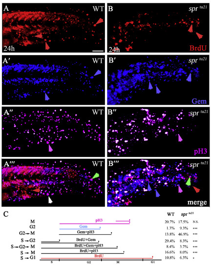
sprtu21 mutant cells spend longer time in G2 and M phases. (A-B‴) The same cells in wild type (A-A‴) and mutant (B-B‴) visualized with different markers to distinguish between the stages of the cell cycle at 24 h. Embryos were labeled for 1 hour with BrdU and then fixed and stained for: anti-BrdU to visualize cells in the S-phase (A, B); anti-GFP to visualize Cerulean-Geminin cells in S/G2/early M phase (A′-B′); and anti-pH3 antibody to visualize cells in mitosis (A″, B″); or the merge (A‴, B‴). Symbols are: red arrows, cells that have gone through a full cell cycle and reside in G1 phase; green arrows, cells that have entered S phase; white arrows, cells that have gone through the S phase and entered mitosis; pink arrows, cells that have entered M phase but did not go through S phase; blue arrows, cells that have entered in the G2/early M phase. Scare bar is 50 μm. (C) Quantification of cells in a specific phase of the cell cycle from the data above. |
| Fish: | |
|---|---|
| Observed In: | |
| Stage: | Prim-5 |
Reprinted from Developmental Biology, 451(2), Petrachkova, T., Wortinger, L.A., Bard, A.J., Singh, J., Warga, R.M., Kane, D.A., Lack of Cyclin B1 in zebrafish causes lengthening of G2 and M phases, 167-179, Copyright (2019) with permission from Elsevier. Full text @ Dev. Biol.