
Fig. 2
- ID
- ZDB-FIG-190820-62
- Publication
- Chapela et al., 2019 - A zebrafish drug screening platform boosts the discovery of novel therapeutics for spinal cord injury in mammals
- Other Figures
- All Figure Page
- Back to All Figure Page
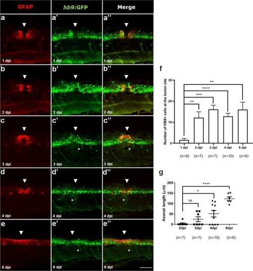
Glial bridge formation and motor neurons regeneration after SCI in 5 dpf larvae. Labeling pattern of Hb9:GFP transgenic larvae with double immunohistochemistry against GFAP (red) to reveal the glial bridge (a–e) and against GFP (green) to reveal HB9+ motor neurons (a’–e’) from 1 to 6 dpi. (a”–e”) Merged channels for each time point. White arrowheads show the lesion site and asterisks highlight the regenerating HB9+ peripheral axons. Rostral side is to the left and dorsal side is up. Number of HB9+ motor neurons (f) and length of HB9+ peripheral axons (g) at the lesion site from 1 to 6 dpi. SCI_spinal cord injury, dpf_days-post-fertilization, hpi_hours-post-injury Mean ± s.e.m. of one experiment is presented. ns - not significant, *p < 0.05, **p < 0.01, ***p < 0.001 and ****p < 0.0001, Student’s t-test with Welch’s correction. Scale bar: 100 µm. |