
Fig. 6
- ID
- ZDB-FIG-190816-32
- Publication
- Fogerty et al., 2019 - Genomic non-redundancy of the mir-183/96/182 cluster and its requirement for hair cell maintenance
- Other Figures
- All Figure Page
- Back to All Figure Page
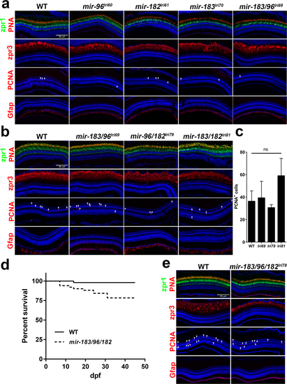
Adult mutants have normal retinal structure, but mir-183/96/182lri78 survival is depressed. (a,b) Survey of retinal anatomy in adult fish at (a) 12 m or (b) 6 m. Markers used here are equivalent to those used for imaging of larval fish, except that we also examined PCNA and Gfap staining to look for evidence of cell proliferation and gliosis, respectively. (c) Quantification of PCNA labeling in double mutants. PCNA + cells were counted across a 10μm thick retinal cross-section at the level of the optic nerve. Proliferating cells in the optic nerve head and in the ciliary marginal zone were excluded. ns = not significant (ANOVA). For all data points, n = 3. (d) Survival curve of fish from incrosses of WT and mir-183/96/182lri78 heterozygous parents. Each of the two cohorts began the study with a population of 51 fry. (e) Histological analysis of a surviving 15wk mir-183/96/182lri78 homozygote, showing normal retinal anatomy. Arrows in (a), (b), and (e) denote PCNA+ nuclei.
|
| Genes: | |
|---|---|
| Antibodies: | |
| Fish: | |
| Anatomical Terms: | |
| Stage: | Adult |
| Fish: | |
|---|---|
| Observed In: | |
| Stage Range: | Days 30-44 to Adult |