Figure 2 - figure supplement 2

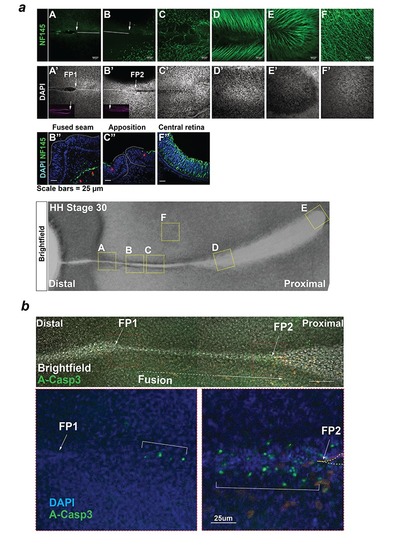
Analysis of axonal processes and apoptosis during chick OFC.

(a) Anti-Neurofilament immunostaining (NF145, Neurofilament medium; Top panels) in the OFM during active fusion. NF145 immunoreactivity (green) was absent from the distal and medial OFM but was observed in the proximal region at the developing pecten and optic nerve. Immunostaining on sections (middle panels) taken at the fused seam, the apposed OFM, and at the central retina confirmed the absence of axons in epithelial tissues at the open OFM and the nascently-fused OFM. Panel labels correspond to positions indicated in the HH.St30 flat-mount brightfield image (Bottom). (b) Top: Combined bright-field and fluorescence confocal imaging (Z-stack projections) of whole mount anti-activated Caspase-3 immunostaining in a HH.St30 fissure showed positive foci in the fused seam at a ~ 100 μm from the static FP1, and multiple positive foci within the fused seam >100 μm FP2. Below: Enlarged images of FP1 and FP2 from (b) counterstained with DAPI highlighted the enrichment for A-Casp3 foci at FP2 (open margin is indicated by hatching).

Expression Data

Expression Detail
Antibody Labeling
Phenotype Data

Phenotype Detail
Acknowledgments
This image is the copyrighted work of the attributed author or publisher, and ZFIN has permission only to display this image to its users. Additional permissions should be obtained from the applicable author or publisher of the image. Full text @ Elife