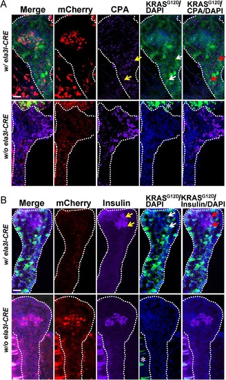
Early KRASG12D-responsive pancreatic progenitors contribute to endocrine as well as exocrine cells. (A) The pancreas from 5 dpf double transgenic Tg (ela3I-CRE; LSL-KRASG12D) larvae was stained with an exocrine specific marker, CPA. CPA staining was observed in the apical cytoplasm as well-developed apical secretory granules (yellow arrows). The GFP signal was detected in both the membrane and cytoplasm as a surrogate marker of KRASG12D activation (white arrows). The merged confocal image showed that the pancreatic progenitor cells expressing oncogenic KRASG12D expressed CPA in apical secretory granules of exocrine pancreas (red arrows). (B) The pancreas from 5 dpf double transgenic Tg (ela3I-CRE; LSL-KRASG12D) larvae was stained with an endocrine specific marker, insulin. Insulin staining was shown in the cytoplasm of islet β-cells (yellow arrows). The merged confocal image showed that some of pancreatic progenitor cells expressing oncogenic KRASG12D co-expressed the endocrine cell marker, insulin (red arrows). Asterisk (*) indicates the auto fluorescence of the gut. Scale bar: 25 µm.
|
