
FIGURE
Fig. 1
- ID
- ZDB-FIG-190723-1086
- Publication
- Gong et al., 2019 - Sec14l3 potentiates VEGFR2 signaling to regulate zebrafish vasculogenesis
- Other Figures
- All Figure Page
- Back to All Figure Page
Fig. 1
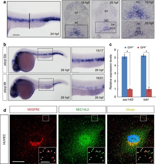
sec14l3/SEC14L2 are expressed in endothelial cells and co-localized with VEGFR2. a Expression pattern of sec14l3 examined by WISH. Trunk vascular expression of sec14l3 is shown at 24 hpf and the vertical line denotes the equivalent position of transverse sections in embryos at 19 hpf and 25 hpf. High-resolution views of the boxed regions are shown at right panels. Adjacent tissues are denoted. NT, neural tube; NC, notochord; DA, dorsal aorta; PCV, posterior cardinal vein; VC, vascular cord. Scale bars, 100 μm. b The absence of sec14l3 expression in the trunk vascular system of etsrp mutant embryos at 26 hpf. High-resolution views of the boxed regions are shown at right panels. Scale bars, 100 μm. c Quantitative RT-PCR of sec14l3 mRNA in both GFP+ cells and GFP- cells from Tg(fli1a:EGFP)y1 transgenic embryos at 24 hpf. sec14l3 is highly enriched in GFP+ cells (blue bars) relative to negative cells (red bars), using β-actin as the internal control. kdrl/vegfr2 mRNA serves as a positive control for vascular endothelial cells. A two-tailed t-test was used for statistical analysis. *p < 0.05, the exact p-values in each figure are shown in the Source data file. d SEC14L2 is co-localized with VEGFR2 in HUVECs. Red and green represent anti-VEGFR2 antibody and anti-SEC14L2 antibody staining signal respectively; blue symbolizes DAPI stained nucleus. Regions in the boxes are enlarged in the right corner. Scale bars, 20 μm in the original pictures and 5 μm in the enlarged panelssec14l3/SEC14L2 are expressed in endothelial cells and co-localized with VEGFR2. a Expression pattern of sec14l3 examined by WISH. Trunk vascular expression of sec14l3 is shown at 24 hpf and the vertical line denotes the equivalent position of transverse sections in embryos at 19 hpf and 25 hpf. High-resolution views of the boxed regions are shown at right panels. Adjacent tissues are denoted. NT, neural tube; NC, notochord; DA, dorsal aorta; PCV, posterior cardinal vein; VC, vascular cord. Scale bars, 100 μm. b The absence of sec14l3 expression in the trunk vascular system of etsrp mutant embryos at 26 hpf. High-resolution views of the boxed regions are shown at right panels. Scale bars, 100 μm. c Quantitative RT-PCR of sec14l3 mRNA in both GFP+ cells and GFP- cells from Tg(fli1a:EGFP)y1 transgenic embryos at 24 hpf. sec14l3 is highly enriched in GFP+ cells (blue bars) relative to negative cells (red bars), using β-actin as the internal control. kdrl/vegfr2 mRNA serves as a positive control for vascular endothelial cells. A two-tailed t-test was used for statistical analysis. *p < 0.05, the exact p-values in each figure are shown in the Source data file. d SEC14L2 is co-localized with VEGFR2 in HUVECs. Red and green represent anti-VEGFR2 antibody and anti-SEC14L2 antibody staining signal respectively; blue symbolizes DAPI stained nucleus. Regions in the boxes are enlarged in the right corner. Scale bars, 20 μm in the original pictures and 5 μm in the enlarged panels |
Expression Data
| Gene: | |
|---|---|
| Fish: | |
| Anatomical Terms: | |
| Stage Range: | 20-25 somites to Prim-5 |
Expression Detail
Antibody Labeling
Phenotype Data
| Fish: | |
|---|---|
| Observed In: | |
| Stage: | Prim-5 |
Phenotype Detail
Acknowledgments
This image is the copyrighted work of the attributed author or publisher, and
ZFIN has permission only to display this image to its users.
Additional permissions should be obtained from the applicable author or publisher of the image.
Full text @ Nat. Commun.