
Fig. 3
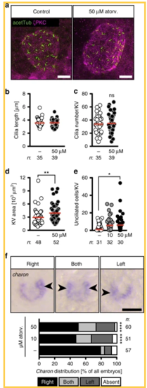
Atorvastatin impacts on the development of the temporal organ of laterality. a Confocal stacks of 6–8 somite stage (ss) Kupffer’s vesicle (KV). Cilia were stained with an antibody against acetylated tubulin (acetTub, green). Apical cell membranes to visualize the KV outline were stained with an antibody against atypical PKC (PKCζ, magenta). Scale bar: 20 µm. b Bar graph displaying the length of KV cilia in control and atorvastatin-treated embryos; n = 1231 (DMSO) and 1424 (50 µM atorv.) cilia; p = 0.6357, two-tailed Mann–Whitney test. c Cilia number per KV was not changed in the presence of atorvastatin; p = 0.6861, two-tailed t-test with Welch’s correction. d KV area was larger upon atorvastatin treatment. Each circle is one KV and embryo. ** p=0.0034. Two-tailed Mann–Whitney test. e Atorvastatin-treated embryos have more unciliated cells in the KV than control embryos. Cilia were stained using anti-acetylated antibody and KV cells were counted using the Sox17-GFP line. KV cells express high levels of GFP and can be distinguished from surrounding cells outside the KV. Each circle symbolizes one embryo; *p = 0.0375, Kruskal–Wallis test with Dunn’s multiple comparison test. f Atorvastatin disturbs correct spatial expression of charon. Representative images of in situ hybridization for charon at 10 ss with correct strong expression on right side and aberrant strong expression on both sides or left side (expression indicated by arrowheads; absent expression not shown). Scale bar: 50 µm. Stacked bar graphs summarises three experiments. Fisher’s exact test; n = 57 (DMSO), 51 (10 µM atorvastatin) and 60 (50 µM atorvastatin); ****p < 0.0001 for DMSO vs 10 µM as well as vs 50 µM. b–f Numbers of embryos analysed indicated below or next to graphs |
| Gene: | |
|---|---|
| Antibodies: | |
| Fish: | |
| Condition: | |
| Anatomical Terms: | |
| Stage Range: | 5-9 somites to 10-13 somites |
| Fish: | |
|---|---|
| Condition: | |
| Observed In: | |
| Stage Range: | 5-9 somites to 10-13 somites |