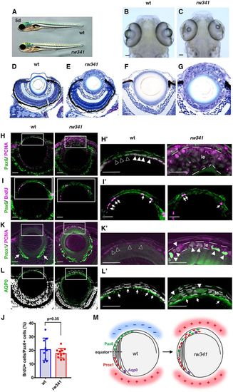
Ectopic lens fiber differentiation occurs in rw341 mutant lens epithelium. (A) Wild-type and rw341 mutant embryos. (B,C) Wild-type (B) and rw341 mutant (C) heads. (D,E) Retinas of wild type (D) and rw341 mutants (E). rw341 mutants show a small lens fiber core, which is surrounded by many aggregated lens cells. (F,G) Higher magnification of the lenses in D and E. (H,H′) Labeling of lenses using anti-Pax6 and PCNA antibodies. In wild type, lens epithelial cells express Pax6 and PCNA (H′, filled arrowheads). Open arrowheads in H′ indicate cells with relatively weak PCNA expression. In multilayered lens epithelium of rw341 mutants (H′, le), most cells express PCNA, but Pax6 expression is weak. (I,I′) Labeling of lenses using anti-Pax6 and BrdU antibodies. Arrows indicate BrdU signals. (J) Percentage of BrdU-positive cells among Pax6-positive lens epithelial cells. Data are mean±s.d. No statistical difference between wild type and rw341 mutants (Student's t-test). (K,K′) Labeling of lenses using anti-Prox1 and PCNA antibodies. In wild type, Prox1 is not expressed in lens epithelium (K′, open arrowheads) and only in early differentiating lens fiber cells (K, arrows). In rw341 mutants, many lens epithelial cells (K′, le) express Prox1 (K′, filled arrowheads). (L,L′) Labeling of lenses using anti-AQP0 antibody. AQP0 is expressed in elongating lens fiber cells in wild type and rw341 mutants (L′, arrows). AQP0-expressing cells were observed in multilayered rw341 mutant lens epithelium (L′, arrowheads). (M) Schematic drawing of wild-type and rw341 mutant lenses. Pax6 is expressed in lens epithelium, whereas Prox1 and AQP0 are expressed in newly differentiating and later elongating lens fiber cells, respectively. In rw341 mutants, ectopic lens fiber differentiation occurs in multilayered lens epithelium. Areas where lens fiber differentiation occurs and does not occur are indicated by red (+) and blue (−), respectively. All lenses are at 5 dpf. Scale bars: 40 µm in B,C; 20 µm in D-L′.
|
