
Fig. S2
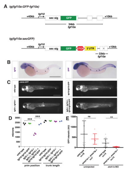
Characterization of the fgf10a:fgf10a-GFP transgenic line, Related to Figure 2. (A) Schematic of fgf10a:fgf10a-GFP and fgf10a:sec-GFP BAC transgenes. White boxes represent exons, and black lines in-between represent introns. STOP represents the stop codon after the GFP coding sequence and 3’UTR indicates the fgf10a 3’UTR fused directly to GFP in exon1. The same fgf10a 3’UTR sequence is also present in exon 3. (B) mRNA in situ hybridization against fgf10a in a wild-type embryo (left) and against GFP in a fgf10a:fgf10a-GFP embryo (right). Arrows indicate expression in neuromasts and arrowhead indicates expression in the primordium. Scale bar = 500 μm. (C) Neuromast and primordium labeled by GFP in cldnb:lyn2GFP embryos of the indicated genotypes at 50 hpf. Arrow indicates position of the primordium. Scale bar = 500 μm. (D) Quantification of the position of the primordium at 50 hpf and trunk length (distance from the posterior edge of the otic vesicle to the front of the primordium or the tip of the tail, respectively) for the indicated genotypes. Black lines represent the mean and data points are individual embryos. *** = p < 0.001. (E) Total Fgf10a-GFP intensity in mature microlumina at the fourth apical constriction from the front in uninjected embryos (left) and embryos injected with atoh1a morpholino of the indicated genotypes (right). Dark black and dark red lines are means and data points are individual embryos. n.s. = p > 0.05, ** = p < 0.01. |
| Genes: | |
|---|---|
| Fish: | |
| Anatomical Terms: | |
| Stage Range: | Prim-15 to Long-pec |
| Fish: | |
|---|---|
| Observed In: | |
| Stage: | Long-pec |
Reprinted from Developmental Cell, 46(6), Wang, J., Yin, Y., Lau, S., Sankaran, J., Rothenberg, E., Wohland, T., Meier-Schellersheim, M., Knaut, H., Anosmin1 Shuttles Fgf to Facilitate Its Diffusion, Increase Its Local Concentration, and Induce Sensory Organs, 751-766.e12, Copyright (2018) with permission from Elsevier. Full text @ Dev. Cell