
Fig. 2
- ID
- ZDB-FIG-180911-1
- Publication
- Messchaert et al., 2018 - Eyes shut homolog is important for the maintenance of photoreceptor morphology and visual function in zebrafish
- Other Figures
- All Figure Page
- Back to All Figure Page
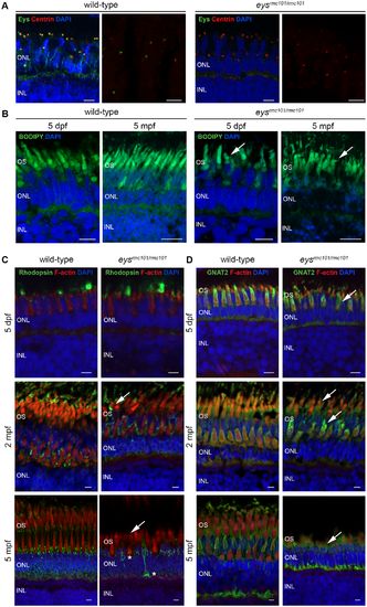
Immunohistochemistry on retinal sections of wild-type and eysrmc101/rmc101 zebrafish. (A) Retinal sections of wild-type and eysrmc101/rmc101 zebrafish at 5 dpf and 5 mpf stained with antibodies against Eys (green) and centrin (red). (B) BODIPY (green) staining showing disorganization of photoreceptor outer segments in eysrmc101/rmc101 zebrafish (5 dpf and 5 mpf) compared to age- and strain-matched wild-type zebrafish (arrows). (C) Retinal sections of wild-type and eysrmc101/rmc101 zebrafish at 5 dpf (upper panel), 2 mpf (middle panel) and 5 mpf (lower panel) stained with antibodies against rhodopsin (green) and F-actin (red). Asterisks indicate mislocalization of rhodopsin to the inner segments and synapses of photoreceptor cells. (D) Retinal sections of wild-type and eysrmc101/rmc101 zebrafish at 5 dpf, 2 mpf and 5 mpf stained with antibodies against GNAT2 (green) and F-actin (red). Arrows indicate dysmorphic outer segments in mutant zebrafish. In all images, nuclei are counterstained with DAPI (blue). INL: inner nuclear layer; ONL: outer nuclear layer; OS: outer segments. Scale: 5 μm. |
| Genes: | |
|---|---|
| Antibodies: | |
| Fish: | |
| Anatomical Terms: | |
| Stage Range: | Day 5 to Adult |
| Fish: | |
|---|---|
| Observed In: | |
| Stage Range: | Day 5 to Adult |