
FIGURE
Fig. S4
- ID
- ZDB-FIG-180911-2
- Publication
- Messchaert et al., 2018 - Eyes shut homolog is important for the maintenance of photoreceptor morphology and visual function in zebrafish
- Other Figures
- All Figure Page
- Back to All Figure Page
Fig. S4
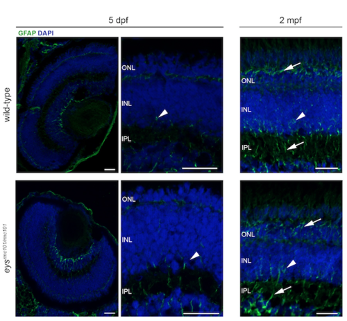
Immunohistochemistry on retinal sections of wild-type and eysrmc101/rmc101 zebrafish in order to investigate Müller glia activation. Retinal sections of wild-type and eysrmc101/rmc101 zebrafish at 5 dpf and 2 mpf stained with antibodies against GFAP (green), as a marker for Müller glia cells. Müller glia cell bodies are located in the inner nuclear layer (arrow heads) and project processes (arrows) in either direction to outer limiting membrane and inner limiting membrane. Nuclei are counterstained with DAPI (blue). INL: inner nuclear layer; IPL: inner plexiform layer; ONL: outer nuclear layer. Scale bar: 20 μm. |
Expression Data
| Gene: | |
|---|---|
| Fish: | |
| Anatomical Term: | |
| Stage Range: | Day 5 to Days 45-89 |
Expression Detail
Antibody Labeling
Phenotype Data
| Fish: | |
|---|---|
| Observed In: | |
| Stage Range: | Day 5 to Days 45-89 |
Phenotype Detail
Acknowledgments
This image is the copyrighted work of the attributed author or publisher, and
ZFIN has permission only to display this image to its users.
Additional permissions should be obtained from the applicable author or publisher of the image.
Full text @ PLoS One