
Fig. S4
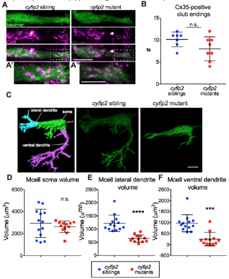
Mauthner cell club endings are normal but dendrite morphology is altered in cyfip2 mutants, Related to Figure 3 (A) Images of Mcell gap junctions labeled by α-connexin 35 (Cx35) Ab (scale bar: 10 μm). Asterisks mark blood cells, dashed boxes highlight magnified region of lateral dendrite with club endings shown in (A’) and (A”) (scale bars: 5 μm). (B) Number of club endings (n=7 siblings, 8 mutants; p=0.11, Mann-Whitney; mean ± SD). (C) Confocal stacks of gap43-citrine-labeled Mcell membranes were masked and rendered in 3D using Imaris. Representative images show severely altered dendritic morphology in cyfip2 mutants (scale bar: 5 μm). (D-E) Quantification of soma, lateral dendrite, and ventral dendrite volume (***p<0.001, ****p<0.0001; Mann-Whitney; mean ± SD). |