
Fig. S7
- ID
- ZDB-FIG-180703-41
- Publication
- Campbell et al., 2018 - Directing Nanoparticle Biodistribution Through Evasion and Exploitation of Stab2-Dependent Nanoparticle Uptake
- Other Figures
- All Figure Page
- Back to All Figure Page
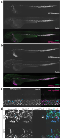
FluoHA colocalization with liposomes. A,B. Whole-embryo view of coinjected fluoHA (green) and A. DOPG liposomes or B. DSPC liposomes (red), 1h after injection, reveals colocalization in PHS, CCV, PCV and CV scavenger endothelial cells. C. Tissue level view of coinjected fluoHA and DOPG liposomes, 1h after injection reveals colocalization in SECs. Monocytes/macrophages (arrowheads) take up DSPC but not fluoHA. D. Cellular view of coinjected fluoHA (green) and DOPG or DSPC liposomes (blue) in mpeg:RFP (red) transgenic embryos. Colocalization of fluoHA with both liposomes is observed in all SECs, but not in macrophages/monocytes, which only take up liposomes, but not fluoHA. |