
Fig. S4
- ID
- ZDB-FIG-180703-38
- Publication
- Campbell et al., 2018 - Directing Nanoparticle Biodistribution Through Evasion and Exploitation of Stab2-Dependent Nanoparticle Uptake
- Other Figures
- All Figure Page
- Back to All Figure Page
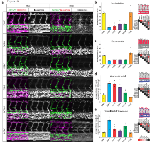
Contribution of individual lipids to liposome biodistribution. A. Cellular view of liposome distribution in kdrl:GFP transgenic embryos, 1h and 8h after injection with liposomes generated from six different individual lipids. B. Quantification of liposome levels in circulation based on rhodamine fluorescence intensity in the lumen of the dorsal aorta at 1h after injection. C. Quantification of extravascular liposome levels based on rhodamine fluorescence intensity outside of the vasculature between the DLAV and DA at 8h after injection. D. Quantification of liposome levels associated with venous vs. arterial endothelial cells based on rhodamine fluorescence intensity associated with caudal vein vs. DA at 8h after injection. E. Quantification of liposome levels associated with the vessel wall based on relative rhodamine fluorescence intensity associated with all endothelial cells vs. rhodamine fluorescence intensity in circulation at 1h after injection. BE. Bar height represents median values, dots represent individual data points, significantly different pairs of values based on Kruskal-Wallis and Dunn’s test with Bonferroni correction are indicated by colored boxes (representing significance levels; CV=critical value; NT=not tested). n=6 individually injected embryos per group (in 2 experiments). |